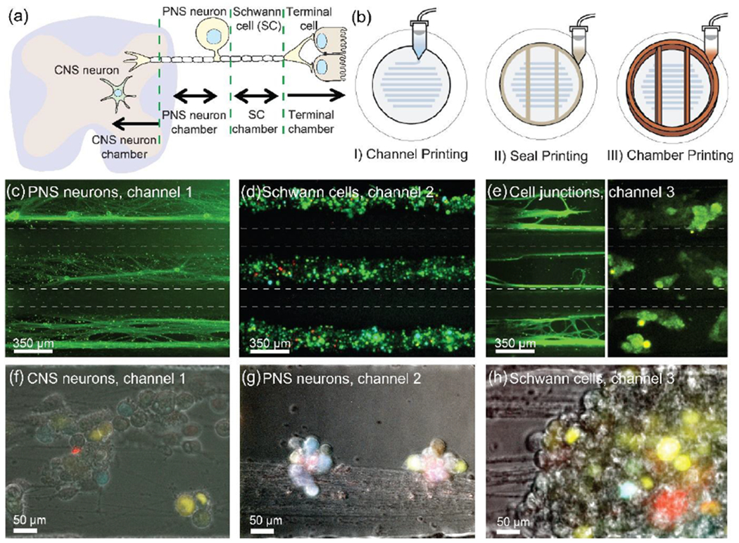
Figure 7.

(a) Schematic of the nervous system showing four primary components: CNS neurons, PNS neurons, Schwann cells, and epithelial cells. (b) 3D printing of tri-chamber consisting of three steps: (I) parallel 350 μm wide channels – providing axonal guidance, (II) a sealant layer – preventing fluidic culture media exchange between the chambers, and (III) a top tri-chamber – providing isolation and organization of specific cell types. (c-e) Biomimetic maturation of 3D printed nervous system on a chip: alignment of axonal networks and spatial organization of cellular components. (c) PNS neurons and axons in chamber 1, (d) Schwann cells in chamber 2, and (e) epithelial cells in chamber 3. (f-h) In vitro model for nervous system viral infection assays: Schwann cells and CNS neurons are resistant to virus infection transmitted from axons (PNS neurons). (f) Infected CNS neurons in chamber 1, (g) infected PNS neurons in chamber 2, and (h) infected Schwann cells in chamber 3 after 10-14 days of culture. Reproduced with permission.[26] Copyright 2016, Royal Society of Chemistry.
