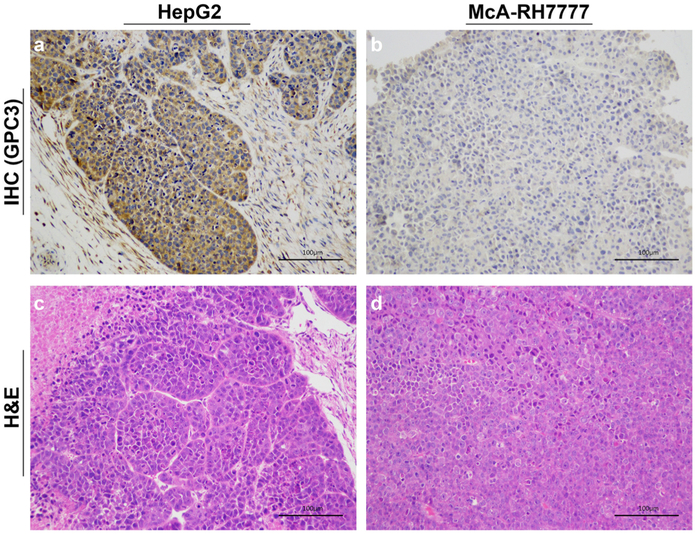
Fig. 6.
Immunohistochemical (IHC) staining of a HepG2 tumor and b McA-RH7777 tumor for GPC3 and hematoxylin and eosin (H&E) staining of c HepG2 tumor and d McA-RH7777 tumor (scale bar: 100 μm). The IHC staining confirmed that the HepG2 tumor is GPC3 positive, and the McA-RH7777 tumor is GPC3 negative.

