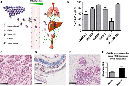
Fig. 1. CXCR4 expression is up-regulated in UM cell lines, hepatic metastases in UM patients, and metastatic UM mice.

(A) Tumor cells that express CXCR4 metastasize through CXCR4-CXCL12 interaction to specific organs that have intrinsically high concentrations of CXCL12 such as the lung, liver, and bone. (B) UM cell lines have elevated CXCR4 expression. Flow cytometry results measured elevated CXCR4 expression across different UM cell lines. Mel290 and M20-09-196 measured more than 80% of CXCR4 immunopositivity. Measurements of each cell line were done in triplicate. (C) CXCR4 IHC staining in liver tissue from metastatic UM patients (n = 4, IRS = 8.2 ± 1.3). The liver metastases displayed strong red intensity, denoting strong CXCR4 expression. (D and E) CXCR4 IHC staining of primary UM (D) and hepatic metastases (E) in metastatic UM mice. UM hepatic metastases have higher CXCR4 expression compared with primary UM, indicated by the red staining. (F) CXCR4 IRS of primary UM and metastases in the liver in metastatic UM mice. Hepatic UM metastases displayed stronger CXCR4 expression (IRS = 9.5 ± 0.8) than primary UM (IRS = 5.4 ± 0.3). P ≤ 0.05.
