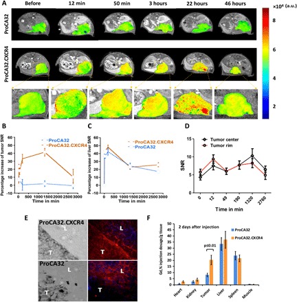
Fig. 5. Progressive MR images of the intrahepatic heterotopic xenotrasplantation UM mice (n = 3 for each group) with Cys-ProCA32.CXCR4 administration.

(A) T1-weighted gradient echo MR images of control mice (with injection of nontargeted agent Lys-ProCA32) and mice with Cys-ProCA32.CXCR4 injection. MRI scans were acquired before and after injection at different time points until 46 hours; tumors are represented by the heat map in MRI images. (B) Percentage increase of SNR of melanoma tumors at different time points shows the dynamic binding process of Cys-ProCA32.CXCR4. For mice that received the Cys-ProCA32.CXCR4 injection, a gradual increase of intensity in melanoma tumor region was observed up to 24 hours, showing the CXCR4-targeting effect, followed by washing out at 46 hours (further time points not acquired). (C) Time plot of the liver SNR percentage increase following Cys-ProCA32.CXCR4 and Lys-ProCA32 injection. The liver SNRs of mice receiving Cys-ProCA32.CXCR4 and Lys-ProCA32 exhibited similar patterns of the SNR time plots, where the liver intensity substantially increased up to 3 hours after injection of both contrast agents, followed by loss of intensity after 3 hours. (D) Time plot of tumor rim and tumor center SNR change of mice with Cys-ProCA32.CXCR4 administration. Cys-ProCA32.CXCR4 exhibited good tumor permeability; tumor rim SNR was enhanced at early time points (12 min after injection). SNR enhancement gradually penetrated to the center of the tumor. At 24 hours after injection, the view of the tumor region following Cys-ProCA32.CXCR4 injection revealed broad distribution and heterogeneous enhancement. (E) Immunofluorescence staining of Cys-ProCA32.CXCR4 and Lys-ProCA32 in the liver (L) and tumor (T) of Mel290 mice. For mice that received Cys-ProCA32.CXCR4 injection (top), Cys-ProCA32.CXCR4 accumulated in the UM tumor tissue (denoted by red fluorescence). For the mice injected with Lys-ProCA32 (bottom), UM tumors exhibited dark fluorescence intensity relative to the UM tumor regions of the mice that received Cys-ProCA32.CXCR4 injection. (F) ICP-OES analysis of Gd3+ tissue distribution 2 days after injection of ProCAs. Mice with Cys-ProCA32.CXCR4 injection exhibited significantly more Gd3+ distribution in tumor tissue than mice that received Lys-ProCA32 injection (P < 0.01).
