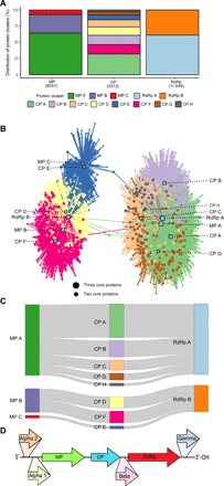
Fig. 2. Examination of ssRNA phage proteins.

(A) Distribution of protein hits (in parentheses) across MP, CP, and RdRp clusters was identified using HMM 5-MC. (B) Bipartite connection network of contigs (circles) with proteins (squares). Colors are based on the associated CP from (A). (C) Protein cluster co-occurring profiles of ssRNA phages having all three full-length core proteins and (D) the frequently observed positions of hypothetical proteins (genes not drawn to scale).
