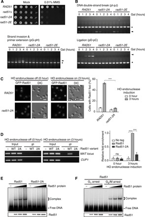
Fig. 3. The G2/M-phase CDK1-dependent phosphorylation promotes the DNA binding affinity of Rad51.

(A) Results from the serial dilution assay used to assess MMS sensitivity of rad51Δ cells expressing Rad51 variants. Cells were spotted in 10-fold serial dilutions on SC medium in the absence or presence of 0.01% MMS. rad51-2A indicates rad51Δ cells expressing the Rad51 mutant with alanine substitutions at Ser125 and Ser375. rad51-2E indicates rad51Δ cells expressing the Rad51 mutant with glutamate substitutions at Ser125 and Ser375. (B) Homologous recombination efficiency test of rad51Δ cells expressing Rad51 variants. Genomic DNA was extracted every 1 hour after 2% galactose addition and analyzed by PCR. Arrowheads indicate the PCR products of the homologous recombination intermediates. Asterisks indicate the PCR products of the control region (ARG5,6). (C) Accumulated GFP-tagged Rad51 and Rad51-2A at the DNA damage sites. To make DSB, HO endonuclease was induced during incubation with 2% galactose for 3 hours. DIC, differential interference contrast. Scale bars, 4 μm. The percentages of cells with Rad51-GFP foci are shown on the right panel. Data are presented as the means ± SD of triplicate experiments. P values were determined by Student’s t test (***P < 0.005). (D) ChIP assay was used to assess the DNA binding affinity of Rad51 and Rad51-2A. HO endonuclease was induced during incubation with 2% galactose for 3 hours. Immunoprecipitated DNA with Rad51 variants was analyzed for the MAT locus by PCR (left panel) and by quantitative real-time PCR (right panel). CUP1 was used as a negative control. Data are presented as the means ± SD of triplicate experiments. P values were determined by a one-sample t test (***P < 0.005). ns, not significant. (E) Results from the EMSA used to assess the ssDNA binding affinity of Rad51 and Rad51-2A. EMSA was performed using a binding buffer that includes 35 mM tris-Cl (pH 7.5), 5 mM ATP, 5 mM MgCl2, 50 mM KCl, bovine serum albumin (100 μg/ml), and 1 mM dithiothreitol. (F) Results from the EMSA used to assess the DNA binding affinity of the G1- and the G2/M-phase Rad51. α-Factor (150 μM) and nocodazole (15 μg ml−1) were treated to synchronize cell cycle to the G1 and G2/M phases, respectively.
