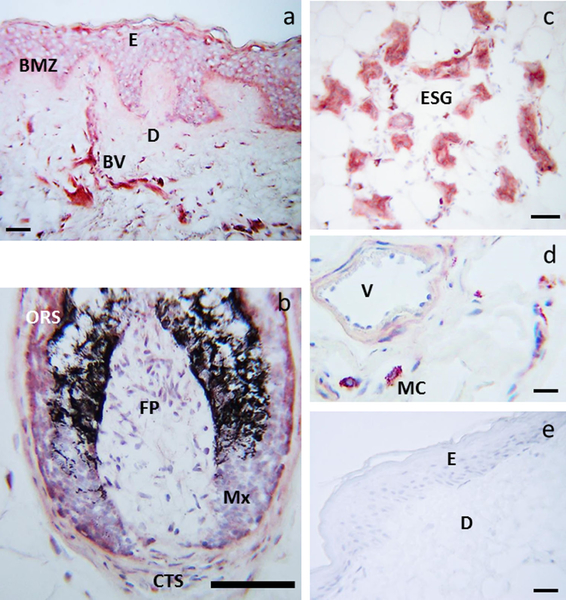
Figure 3.
Immunolocalization of serotonin in human scalp. Serotonin immunoreactivity was detected cytoplasmically and membranously in keratinocytes and melanocytes of the basal layer, and in keratinocytes of the suprabasal and spinous layers, as well as in dermal blood vessels (BV) (a), ORS and matrix cells of the hair follicle, FP and CTS (b), ESG (c) and mast cells (d). Control: primary antibody replaced with donkey serum and secondary antibody only (e). BMZ: basement membrane zone, CTS: D: dermis, E: epidermis, ESG: eccrine/sweat glands, FP: dermal papilla fibroblasts, Mx: hair follicle matrix, ORS: outer rout sheath, V: lumen of the blood vessel. Scale bar: 20 μm (a), 100 μm (b), 50 μm (c), 13 μm (d), 23 μm (e). The immunostains are from the scalp skin of the 35 years old male donor, a representative of three donors with multiple stains performed for each skin sample

