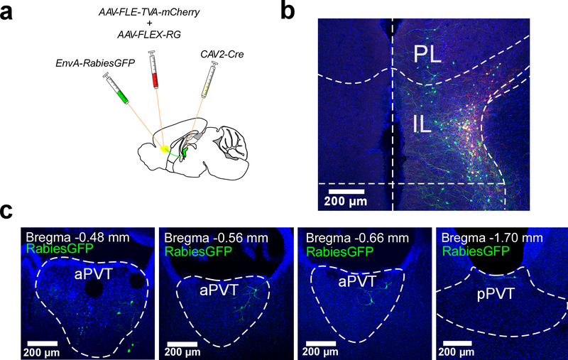
Extended Data Fig. 5. Pseudo-typed rabies tracing of monosynaptic inputs onto ILPVT neurons.
a. Schematic of the viral vector strategy employed for the pseudo-typed rabies tracing of monosynaptic inputs to ILPVT neurons. b. Representative image of Rabies-GFP and TVA-mCherry double-labelled cells illustrate the location of rabies starter cells. c. Rabies-GFP (retrogradely-labelled) neurons are observed throughout the aPVT, which is enriched with Type II PVT neurons (Fig. 1). Rabies-GFP neurons were rarely observed in the pPVT and restricted to the most dorsal part of the pPVT, a region of the pPVT dominated by Type II PVT neurons. These findings demonstrate that IL-projection neurons of the PVT are monosynaptically connected to ILPVT neurons, highlighting the presence of a PVT-IL thalamo-corticothalamic loop (data from 2 mice). This experiment was independently repeated twice with similar results.

