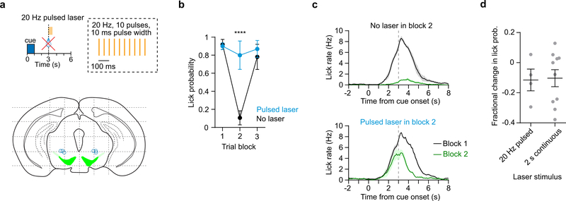
Extended Data Fig. 10: Similar effect of pulsed and continuous laser stimulation during reward extinction.
a. (Top) Illustration of the pulsed laser stimulation (as opposed to 2 s continuous used in Fig. 6) protocol used to activate DA neurons during reward extinction. (Bottom) Histologically determined optical fiber tracks for the 4 Chrimson+ mice targeting the VTA. Grid lines are spaced 1 mm apart.
b. Anticipatory lick probability in each of the three trial blocks on extinction sessions with laser (blue) and without laser (black) (n = 4 Chrimson+ mice, two-way RM ANOVA, group effect: F1,3 = 30.2, P = 0.01, trial block effect: F2,6 = 40.9, P = 0.0003. Post-hoc Sidak’s test: ****P < 0.0001.
c. Mean lick rate versus time for Chrimson expressing animals undergoing reward extinction on extinction sessions without laser (top) and with pulsed laser stimulation (bottom) (n = 4 mice). Black and green lines represent trial blocks 1 and 2, respectively.
d. Fractional change in anticipatory lick probability during reward extinction experiments with 2 s continuous (n = 10 mice) and 20 Hz pulsed laser (n = 4 mice). There is no significant difference between these groups (two-sided unpaired t-test, t12 = 0.1, P = 0.91). Data are expressed as mean ± SEM.

