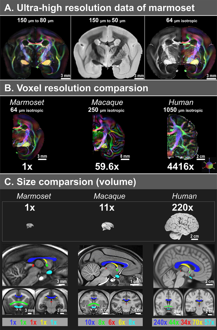
Figure 1. Comparison of isotropic data resolution, brain volume, and fiber volumes between marmosets, macaques, and humans.
(A) Compared to our previous 150 μm data16, the new dataset has a substantial improvement in spatial resolution, including 80 μm dMRI (left), 50 μm T2* (middle), and 64 μm dMRI (right). (B) Comparison of voxel resolution. The macaque ex-vivo dMRI dataset has 250 μm resolution20, and the human in-vivo dMRI dataset from the 7T human connectome project21 has 1050 μm resolution. In terms of volume (μm3), the resolution of our 64 μm data is 59.6x and 4416x higher than that of the macaque and human data, respectively. (C) Comparison of the brain and fiber volumes (μm3). Brain sample images are from brainmuseum.org. All volumes are estimated from three population-based templates, including a marmoset template from 22 adult marmosets, a NMT macaque template39, and a MNI152 adult human template40. The five white matter tracts shown are the corpus callosum (blue), the anterior commissure (green), the posterior commissure (red), the fornix (yellow), and the optic white matter (cyan; including the optic nerve, the optic chiasm, and the optic tract).

