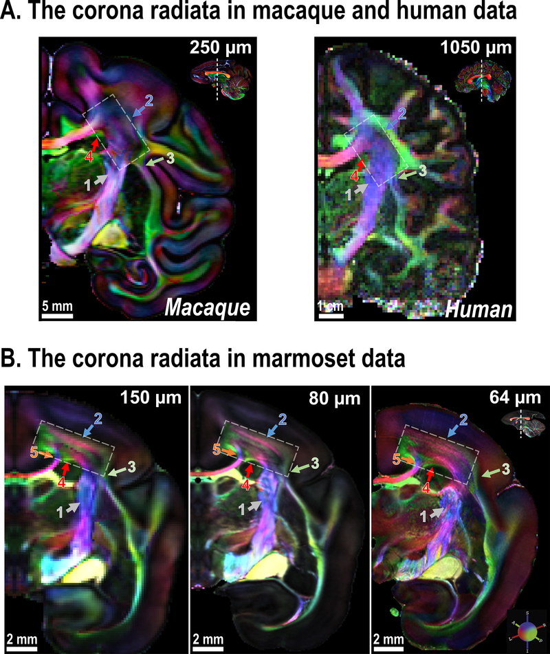
Figure 2. The corona radiata imaged at different spatial resolutions.
(A) Coronal slices of the corona radiata (white rectangle) of an ex-vivo macaque brain (at 250 μm) and an in-vivo human brain (at 1050 μm). Note that the corona radiata is mostly dominated by projection fibers from the internal capsule (ic; gray arrow-1). (B) Coronal slices that show the corona radiata of marmoset brain acquired at increasing spatial resolution. Colored arrows highlight areas that benefit from the use of higher spatial resolution (see text for details; the zoomed-in view is provided in Supplementary Fig. 1).

