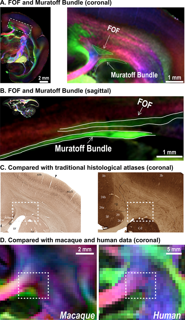
Figure 3. Fronto-occipital fasciculus (FOF) and the Muratoff bundle.
(A) Coronal slice (64 μm) through the corona radiata of the marmoset, where the FOF and the Muratoff bundle are highlighted. (B) Sagittal slice (80 μm) showing the FOF and Muratoff bundle. Two white matter fiber pathways run in a similar direction (green: anterior-posterior) and are separated by a small fiber running left-to-right (red). (C) Non-phosphorylated neurofilament staining of coronal slices from the Paxinos atlas41 and the Hardman atlas42 (modified with permission from the original authors) show that previous histological marmoset atlases cannot distinguish the two fiber bundles due to the lack of fiber-orientation contrasts. (D) As well, the macaque (250 μm) and human dMRI data (1050 μm) cannot distinguish the two fiber bundles due to their insufficient spatial resolution.

