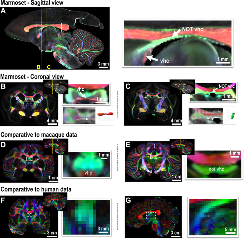
Figure 5. The ventral hippocampal commissure (vhc) of primates.
(A) Middle sagittal slice of the FA-weighted DEC image of the marmoset brain (80 μm). White arrows highlight the vhc and the area mistakenly labeled as the vhc in most atlases. (B) A coronal slice of the marmoset that shows dominant left-right fiber orientation in the vhc. The bottom right is the fiber orientation distribution image for each voxel and overlaid on the FA image, and the fiber orientation of the voxel highlighted by the white dot is presented. (C) A coronal slice of the marmoset brain showing the non-vhc region (fornix area), in which fiber orientation is dominated by the anterior-posterior (DEC-green) direction. (D-E) The vhc and non-vhc white matter in the macaque brain, which shows a relatively smaller vhc. (F-G) The vhc of the human brain is too small to be identified by the in-vivo connectome data (1050 μm). A coronal image is shown in F, and a middle sagittal image is shown in G.

