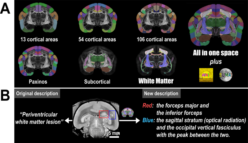
Figure 6. Atlas version 2 and its applications in preclinical research.
(A) Version 2 of the Marmoset Brain Mapping Atlas includes not only the new white matter atlas but also atlases of different cortical parcellation and subcortical gray matter structures from version 116. All these atlases are provided in the same coordinate space at 80 μm and 50 μm with multi-modal MRI templates. These atlases and high-resolution templates are integrated into the AFNI software to provide a fully featured atlas utility. (B) An example from a marmoset disease model that demonstrates how our atlas can improve the localization of the white matter lesion. The original description is cited from29.

