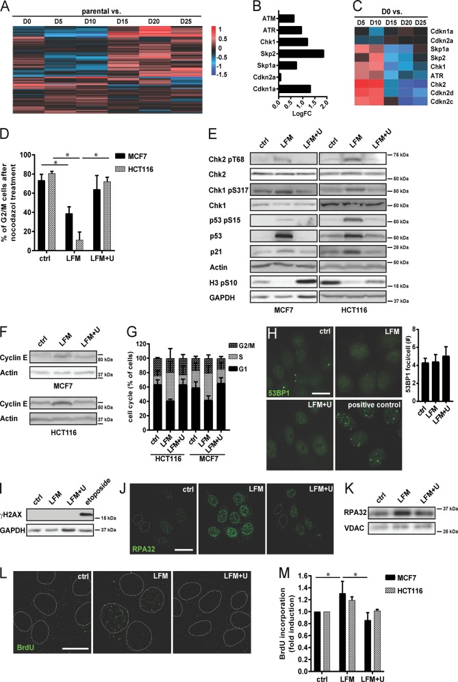
Fig. 1. DHODH inhibition induces replication stress and cell cycle arrest.
a–c Parental 4T1 cells and D0–D25 sublines were subjected to microchip analysis and heat map for the 194 transcripts involved in cell cycle and p53 signaling pathway was constructed. d–m MCF7 and HCT116 cells were exposed to leflunomide (LFM; 50 µM) for 72 h in the presence or absence of uridine (U; 50 µg/l). d Cells were incubated with nocodazole (10 µM, o/n) and the G2/M fraction was detected by flow cytometry using propidium iodide (2.5 µg/ml). e Immunoblotting detection of Chk2 pT68, Chk2, Chk1 pS317, Chk1, p53 pS15, p53, p21, and H3 pS10 and f cyclin E protein level was determined. β-Actin and GAPDH were used as a loading control. g MCF7 and HCT116 cells were assessed for cell cycle distribution. h Immunofluorescent detection of 53BP1 in MCF7 cells was performed. MCF7 cells treated with Etoposide (10 μM) for 24 h were used as a positive control. Scale bar, 10 µm. i Immunoblotting detection of γH2AX signal was performed in MCF7 cells. GAPDH was used as a loading control. j Accumulation of the RPA32 protein on DNA in MCF7 cells was evaluated by immunofluorescence staining and k by immunoblot analysis. VDAC was used as a loading control. l Accumulation of BrdU staining on non-denaturated ssDNA in MCF7 cells was evaluated by immunofluorescence staining and m by FACS. In d, g and m, data are shown as mean ± SEM, n = 3. *P < 0.05, two-way ANOVA. In other panels, representative experiment (from a total number of three experiments) is shown.

