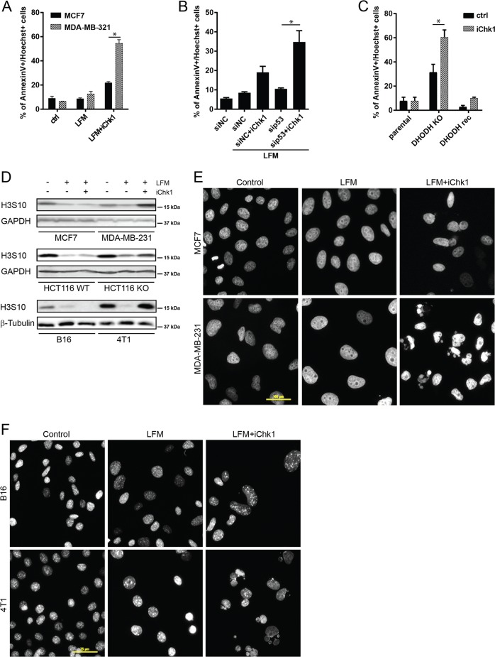
Fig. 5. Inhibition of Chk1 promotes cell death in DHODH-inhibited p53-deficient tumor cells.
a MCF7 and MDA-MB-321 cells were pre-treated with Chk1 kinase inhibitor LY2603618 (iChk1, 5 µM, 30 min before LFM) followed by LFM treatment (50 µM, 48 h) and cell death was evaluated by annexin V/Hoechst staining using flow cytometry. b MCF7 cells were pre-treated with iChk1 (5 µM, 30 min before LFM) after downregulation of p53 using specific siRNA followed by LFM treatment (50 µM, 48 h). Cell death was detected by annexin V/Hoechst staining using FACS. c 4T1 parental, DHODH knock-out (KO), and DHODH reconstituted (rec) cells were treated with iChk1 (5 µM, 48 h) and cell death was evaluated by annexin V/Hoechst staining using FACS. d–f MCF7, MDA-MB-321, HCT116 wt p53, HCT116 p53KO, B16, and 4T1 cells were pre-treated with iChk1 (5 µM, 30 min before LFM) followed by LFM treatment (50 µM, 24 h). d Protein level of H3 pS10 was detected by immunoblot. GAPDH or β-tubulin was used as a loading control. e, f Nuclear morphology was detected by a fluorescent microscope using DAPI staining. Scale bar represents 100 µm (e) or 25 µm (f). In a–c, data are shown as mean ± SEM, n = 3. *P < 0.05, two-way ANOVA. In other panels, representative experiment (from total number of three experiments) is shown.

