Fig. 2.
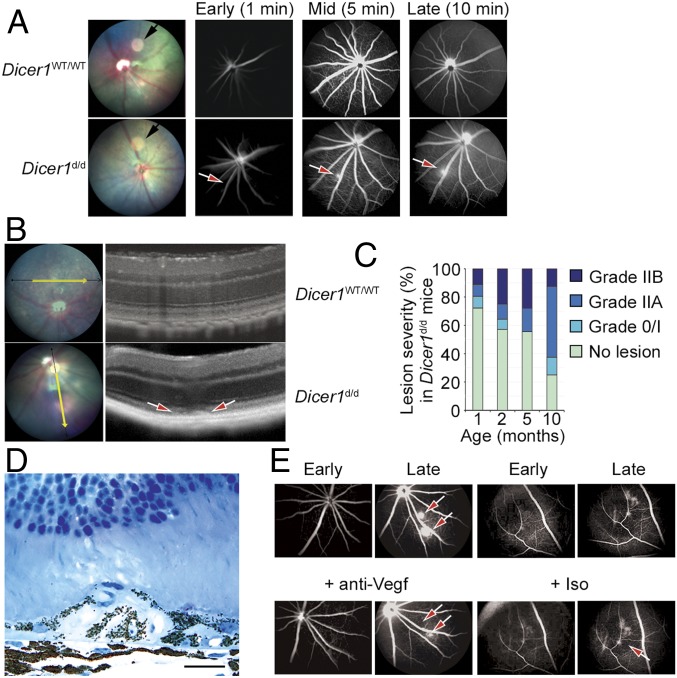
(A) Fundus retinal imaging (Left) and early, mid, and late fluorescein angiograms of wild-type (WT) littermate and Dicer1d/d mice. The black arrow in fundus retinal image denotes circular image artifact. The red arrow denotes focal hyperfluorescent neovascular lesion. (B) Image-guided SD-OCT of a WT littermate (Top) and Dicer1d/d mouse eye (Bottom). The red arrows denote neovascular lesion. (C) Incidence and severity of neovascular lesions Dicer1d/d with respect to age. Ninety individual examinations were included in this analysis. No vascular lesions were detected in WT littermate mice at any age. Age was significantly associated with incidence and severity of neovascular lesions by linear regression (P = 0.0184) and Spearman’s rank (P < 0.00058), respectively. (D) High-magnification toluidine blue-stained 1-μm-thick section of a neovascular lesion in a 12-mo-old Dicer1d/d mouse shows RPE delamination and migration. Scale bar, 20 microns. (E) Representative early and late fluorescein angiograms of Dicer1d/d mouse prior to and 3 d after intravitreous injection of Vegf neutralizing antibody or isotype. The red arrows denote neovascular lesion that resolved following Vegf neutralization.

