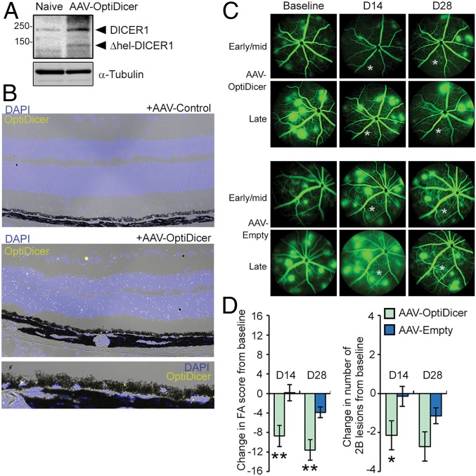
Fig. 8.
Detection of Δhel-DICER1 in retina following subretinal injection of AAV-OptiDicer by immunoblotting (A) and in situ hybridization using a probe antisense to the synthetic OptiDicer sequence (B). (C) Representative fluorescein angiograms of JR5558 mice prior to, and 14 and 28 d after subretinal injection of AAV-OptiDicer or AAV-Empty. Injections were made in an area encompassing the lower left quadrant of the fundus relative to the optic nerve. Approximate injection site is denoted by an asterisk (*). (D) Quantification of changes in total FA score and number of 2B lesions from baseline after AAV-OptiDicer- and AAV-Empty-injected eyes (n = 7 eyes/treatment). *P < 0.05; **P < 0.01.

