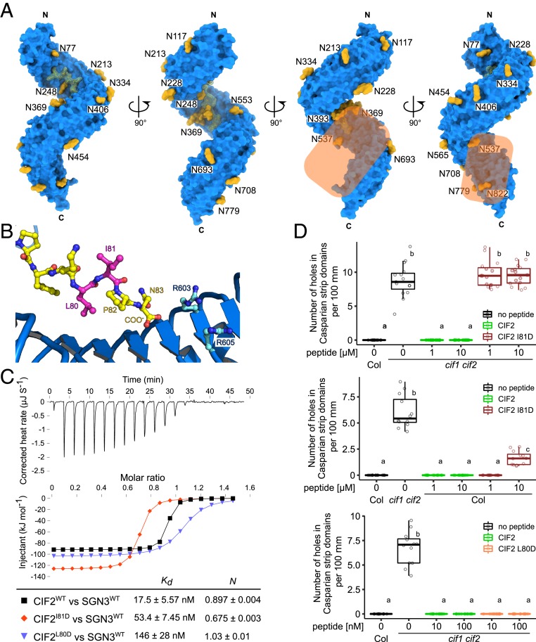
Fig. 6.
Structural and biochemical evidence for a coreceptor kinase required for GSO1/SGN3 activation. (A) The GSO1/SGN3–CIF complex structure reveals a potential coreceptor binding site. Shown is the GSO1/SGN3 ectodomain (surface representation, in blue) in complex with the CIF2 peptide (surface view and bonds representation, in yellow), N-glycans (surface representation in yellow). The potential coreceptor binding surface not masked by carbohydrate is highlighted in orange. (B) Close-up view of CIF2 C terminus bound the GSO1/SGN3, indicating the positions of the side chains of Leu80 (pointing toward the receptor) and Ile81 (pointing to the solvent) (in magenta). (C) ITC assays of CIF2 mutant peptides versus the SGN3 wild-type ectodomain. (D) Quantitative analyses of number of holes in Casparian strip domains per 100 µm in cif1 cif2 double mutants upon treatment with CIF2 peptide variants (n = 15 for the Top, n = 12 for the Middle, and n ≥ 11 for the Bottom). Shown are box plots spanning the first to third quartiles, with the bold line representing the median, and circles indicating the raw data. Whiskers indicate maximum and minimum values, except outliers (b and c, statistically significant difference from a, with P < 0.05, one-way ANOVA and Tukey test).

