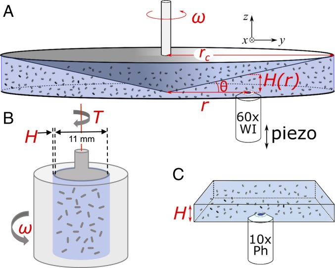
Fig. 2.
Schematic of the three experimental setups used (not to scale). (A) Rheo-imaging setup using cone-plate geometry for visualization during shear. (B) Couette cell for bulk rheometry, after ref. 13. (C) Phase contrast imaging without applied shear.

