Fig. 1.
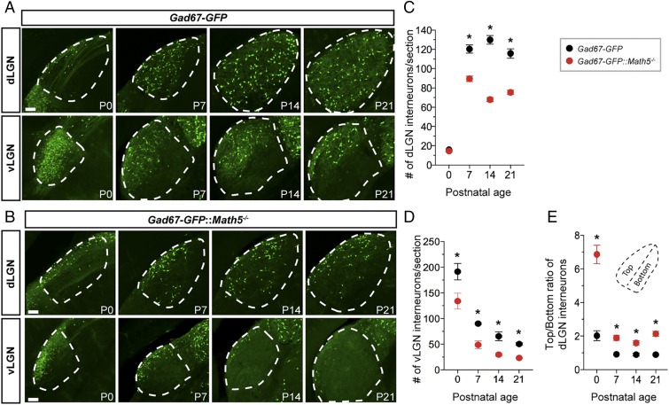
Retinal inputs are necessary for interneuron recruitment into the visual thalamus. (A and B) GFP+ GABAergic interneurons in visual thalamus of P0 to P21 Gad67-GFP (A) and Gad67-GFP::Math5−/− mice (B). The dLGN and vLGN are highlighted by dashed lines. (Scale bars, 70 µm.) (C and D) Summary of age-related changes in GFP+ interneuron number in the dLGN (C) and vLGN (D) of Gad67-GFP and Gad67-GFP::Math5−/− mice. Data points represent means ± SEM, Asterisks (*) represent significance (P < 0.001) between control and mutant [two-way ANOVA, F(9, 178)= 198.2, P < 0.001; Holm–Sidak post hoc test, all P values < 0.0001]. (E) Quantification of interneuron distribution within dLGN of Gad67-GFP and Gad67-GFP::Math−/− mice. Dashed line in inset shows the axis used to delineate the dorsolateral half (“Top”) of dLGN from the ventromedial shell (“Bottom”). Data points represent means ± SEM, Asterisks (*) represent significance (P < 0.001) between control and mutant [two-way ANOVA, F(9, 181) = 53.29, P < 0.0001; Holm–Sidak post hoc test, all P values < 0.0001].

