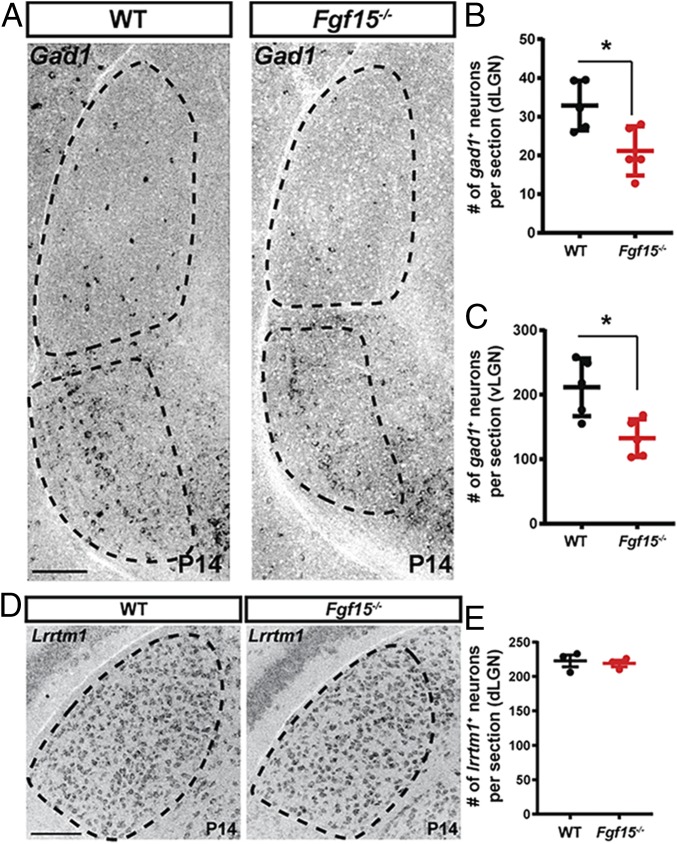
Fig. 5.
Reduced numbers of Gad1+ cells in visual thalamus in the absence of functional FGF15. (A) ISH for Gad1 mRNA revealed reduced numbers of Gad1+ cells in the dLGN and vLGN of P14 Fgf15−/− mutants compared with littermate controls. (B and C) Quantification of Gad1+ cells in the dLGN (B) and vLGN (C) of P14 Fgf15−/− mutants compared with littermate controls (WT). Bars represent means ± SEM. Asterisks (*) represent significantly decreased expression in Math5−/− mutants compared to WT controls by Student’s t-test (P < 0.01). (D) ISH for Lrrtm1 mRNA revealed a normal distribution of relay cells in the dLGN and vLGN of P14 Fgf15−/− mutants compared with littermate controls. (E) Quantification of Lrrtm1+ cells in dLGN of P14 Fgf15−/− mutants compared with littermate controls (WT). Bars represent means ± SEM. (Scale bars, 100 µm for A and D.)

