Fig. 1.
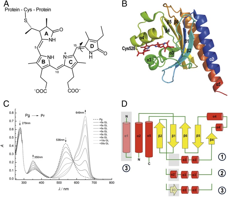
Chromophore structure and absorption spectra. (A) Chemical structure of phycocyanobilin chromophore of Slr1393g3. The chromophore is bound covalently to the protein via a thioether bond between Cys528 and the 31 position of PCB. The molecule is shown in the parental-state configuration (Z,Z,Z,s,s,a). The double-bond photoisomerization (double bond between rings C and D) is indicated by an arrow. (B) Cartoon representation of Slr1393g3 structure in its in vitro-assembled parental Pr state (PDB ID code 5DFY). Secondary-structure elements are labeled according to the AnPixJg2 structure (PDB ID code 3W2Z). The PCB chromophore is shown in stick representation; the covalent bond to Cys528 is highlighted. (C) Illumination-induced conversion between the red- (Pr) and green-absorbing (Pg) forms of Slr1393g3. Formation of the red-absorbing form follows stepwise irradiation of Pg (total irradiation time, 80 s; irradiation source, 670-nm LED). (D) Topology of Slr1393g3 in comparison to AnPixJg2: ① Slr1393g3-Pr state, ② Slr1393g3-Pg and the hybrid form, and ③ AnPixJg2 topology. The topology depicts Slr1393g3 in the parental, red-absorbing state. The gray box between β2 and α3 (part of an unstructured loop) converts into a short helical element in the photoproduct and in the photoisomerization hybrid (coined α2′ in the main text). In the parental state of AnPixJg2, this adapts a β-sheet conformation (β3).

