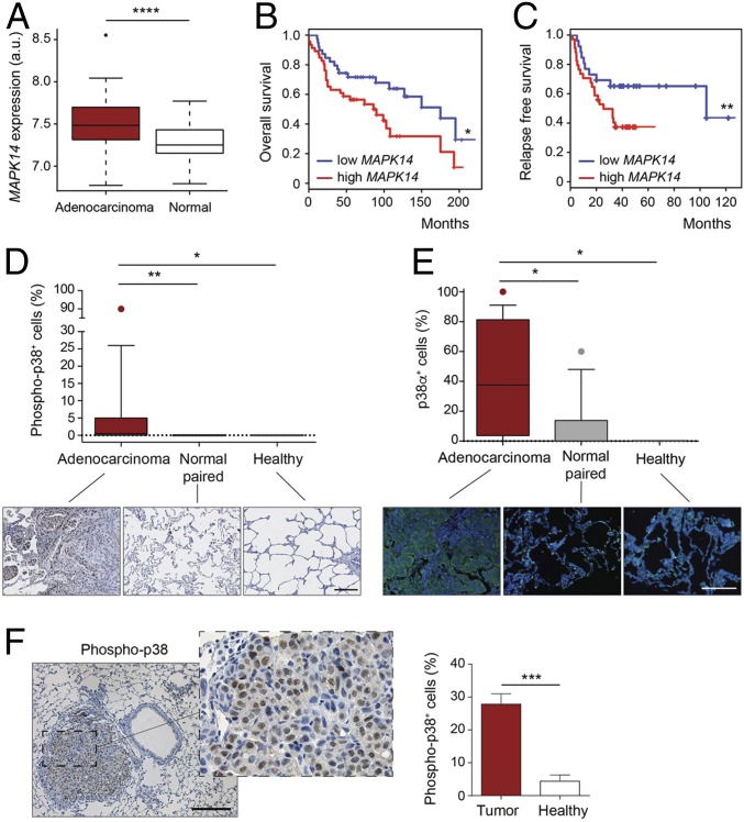
Fig. 1.
High levels of p38α expression correlate with malignancy and poor prognosis. (A) Boxplots for expression levels of MAPK14 mRNA comparing paired adenocarcinomas and healthy tissues (n ≥ 49 per group). a.u., arbitrary units. (B) Kaplan–Meier plots showing a univariate analysis of overall survival of lung adenocarcinoma patients stratified by high or low MAPK14 mRNA expression (n ≥ 39 per group). (C) Kaplan–Meier plots displaying a recurrence-free survival over time of lung adenocarcinoma patients stratified by high or low MAPK14 mRNA expression (n ≥ 26 per group). (D) Representative images and boxplot of the percentage of phospho-p38+ cells in lung adenocarcinoma tumors (n = 17), paired normal tissue (n = 14), and healthy lung parenchyma (n = 5) from a human tissue array. (Scale bar, 200 μm.) (E) The same samples as in D were stained with a p38α antibody by immunofluorescence. DAPI marks nuclei. (F) Representative image of a murine KrasG12V-driven lung tumor stained with phospho-p38 antibody. (Scale bar, 200 μm.) The histogram shows the percentage of phospho-p38+ cells in tumors and in healthy lung parenchyma (n = 5 healthy tissues and n = 21 tumors from 6 different mice). Data represent average ± SEM. *P < 0.05, **P < 0.01, ***P < 0.001, ****P < 0.0001.

