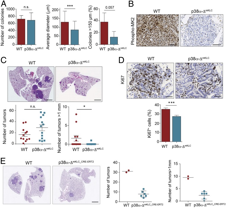
Fig. 5.
Epithelial p38α is necessary for the proliferation of lung tumor cells. (A) WT and p38α-ΔmKLC cells derived from mouse lung tumors were seeded in soft agar. Histograms show the number of colonies formed, their average diameter, and the percentage of colonies bigger than 150 μm per well (n ≥ 42 colonies analyzed). Data represent mean ± SD. (B) Representative images of phospho-MK2 immunostainings of lung tumors formed by intratracheal inoculation of WT and p38α-ΔmKLC cells. (Scale bars, 100 μm.) (C) Representative images of H&E stained lungs from WT animals that were intratracheally inoculated with either WT or p38α-ΔmKLC cells. (Scale bars, 2 mm.) Dot plots show the average number of total tumors and the number of tumors with a diameter bigger than 1 mm at 22 d after the intratracheal inoculation (n ≥ 12 mice per group). Data represent average ± SEM. (D) Representative images of lung tumors from mice that were intratracheally inoculated with either WT or p38α-ΔmKLC cells and stained with Ki67 antibody. (Scale bars, 100 μm.) The histogram shows the quantification of Ki67+ cells per tumor as a percentage of the total number of cells counted (n = 55 WT and n = 70 p38α-ΔmKLC tumors each from ≥5 mice). Data represent average ± SEM. (E) Representative examples of lungs from mice that were intratracheally inoculated with either WT or p38α-ΔmKLC_CRE-ERT2 cells and, 22 d later, treated with tamoxifen. Lungs were stained with H&E 15 d after p38α down-regulation. (Scale bar, 2 mm.) Dot plots show the quantification of both the tumor number and the number of tumors bigger than 1 mm in diameter per animal (n = 2 to 6 mice). *P < 0.05, ***P < 0.001. n.s., not significant.

