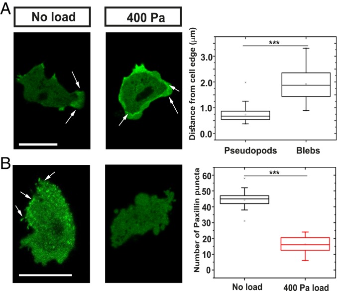
Fig. 3.
Uniaxial load causes cytoskeletal reorganization. (A) Coronin, an F-actin binding protein, relocates under load from pseudopods to the actin scars left behind by blebs. Quantification of the coronin localization from the cell edge. Data are represented as mean ± SD for n ≥ 40 cells for each case; one-way ANOVA, ***P < 0.005. (B) Paxillin patches, thought to mediate adhesion to the substratum, disperse under load. Quantification of the number of paxillin patches in the cell under different loading conditions. Data are represented as mean ± SD for n ≥ 20 cells for each case; one-way ANOVA, ***P < 0.005. Load was applied to aggregation-competent Ax2 cells expressing either coronin–GFP or GFP–paxillin and migrating toward cyclic AMP, under an overlay of 0.5% agarose. (Scale bar: 10 µm.)

