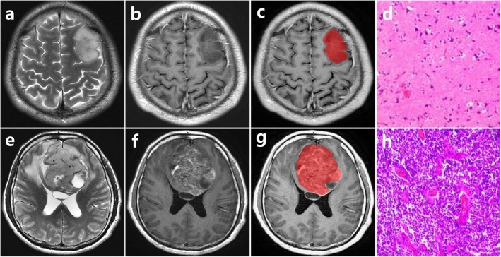
Fig. 7.
Upper row: ODG2 in the left frontal lobe from 33-year-old man; lower row: ODG3 in the bilateral frontal lobe from 46-year-old man. a, e T2-weighted image. b, f T1-weighted contrast-enhanced image. c, g The volume of interest of manually drawn. d, h Pathology slice images show cell density and vascular proliferation

