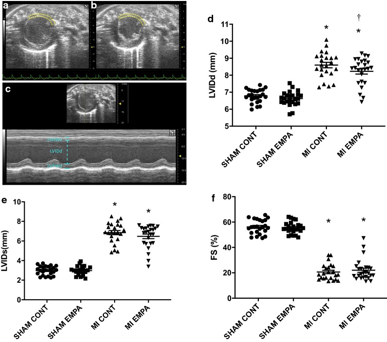
Fig. 1.
Echocardiographic parameters: Representative parasternal short axis views of the LV (Bmode) in diastole (a) and systole (b) 1-week post LAD ligation. The anterolateral infarct is outlined with dotted yellow lines. M-mode imaging (c) reveals thinning of the left ventricular anterior wall (LVAWd), increased internal diameter (LVIDd) and reduced function. d–f represent quantitation of LV internal diameter in diastole, LV internal diameter in systole and fractional shortening. MI reduced FS and led to ventricular dilatation. LVIDd was reduced by empagliflozin therapy. *p < 0.05 MI versus sham, †p < 0.05 versus MI control

