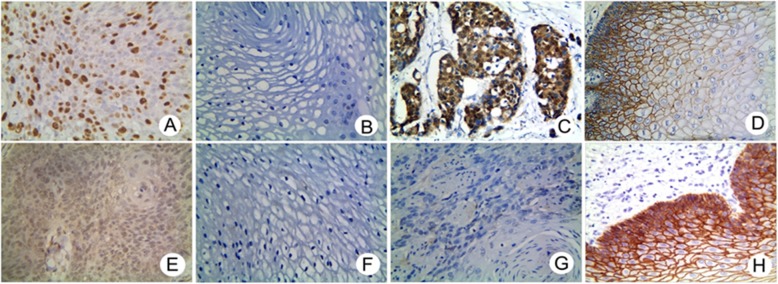
Fig. 2.
Immunostaining of FoxM1, β-catenin, TCF4 and E-cad in ESCC and control tissues (X 400 magnification). a Positive staining of FoxM1 in the nucleus and cytoplasm of tumor cells. b Negative staining of FoxM1 in control tissues. c Positive staining of β-catenin in the nucleus and cytoplasm of tumor cells. d Negative staining of β-catenin in control tissues. e Positive staining of TCF4 in the nucleus of tumor cells. f Negative staining of TCF4 in control tissues. g Negative staining of E-cadherin in tumor cells. h Positive staining of E-cadherin in the membrane and cytoplasm of control tissues

