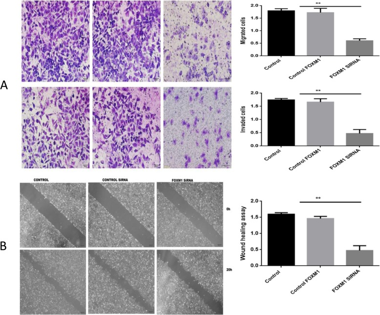
Fig. 6.
a Top panel: Migration and invasion assays following FoxM1 silencing. Bottom panel: Quantitative results are shown for panel. * *P<0.01 vs control (× 200); b Top panel: Wound healing assays to measure the migratory capacity in three groups of EC cells after FoxM1 silencing. Bottom panel: quantitative analysis. **P<0.01 vs control

