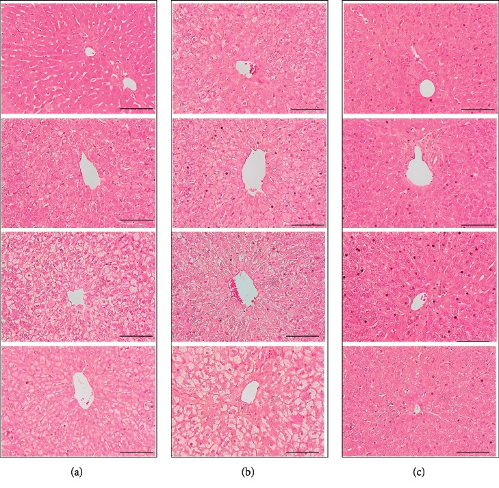
Figure 3.
Histological changes in the livers of cholesterol-fed rabbits by H&E staining. Under higher magnification, hepatocyte ballooning and steatosis were observed throughout the sections in the rabbits from the sham and VO groups. H&E staining showed that steatohepatitis was attenuated in rabbits of LZ8 L. lactis treatment group. CV, central vein. Scale bar = 100 μm. Magnification ×400. (a) Sham group.(b) VO group. (c) LZ8 group.

