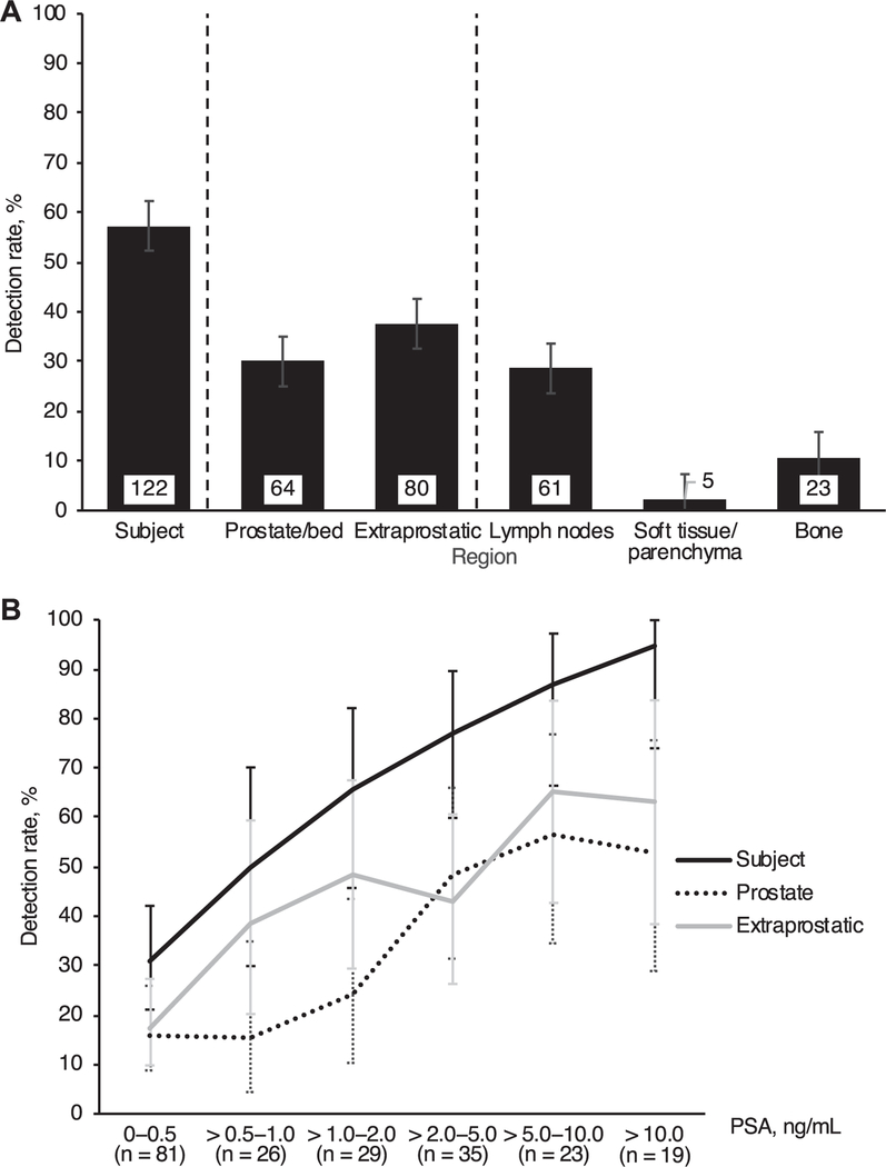
Figure 3.
18F-fluciclovine imaging detection rate by region (A) and PSA (B). Extraprostatic region consisted of lymph nodes, soft tissues/ parenchyma and bone. Lymph nodes consisted of pelvic and extrapelvic (retroperitoneal and other) nodes with overall 24% and 12% detection rate, respectively. Soft tissue/parenchyma positivity was identified in bowel and lung. Error bars indicate 95% CI.

