Figure 5.
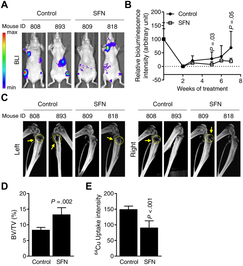
Oral administration of sulforaphane (SFN) inhibits breast cancer-induced osteolytic bone resorption in vivo. A, Representative bioluminescence images of control (corn oil) and SFN-treated (1 mg SFN/mouse in corn oil) mice at 7 weeks after intracardiac injection of MDA-MB-231-Luc cells. Bar at left end indicates intensity of bioluminescence (red representing high intensity and blue representing low intensity). B, Quantitation of bioluminescence of control- (n = 7~8) and SFN-treated (n = 6~9) mice over time. Results shown are mean ± SD. Statistical significance of difference was analyzed by unpaired Student’s t test. C, Representative computed tomography images of affected bones from control- and SFN-treated mice. Yellow dotted circles and arrows indicate bone resorption. D, Quantitation of bone volume (BV)/total volume (TV) of affected hind limbs of control- and SFN-treated mice. Results shown are mean ± SD (n=5 for both groups). Statistical significance of difference was analyzed by unpaired Student’s t test. E, Quantitative plot of ex vivo 64Cu-RGD uptake intensity by affected bones of control- and SFN-treated mice. Results shown are mean ± SD (n=6–7). Statistical significance of difference analyzed by unpaired Student’s t test (n = 7 for control and n = 6 for SFN group).

