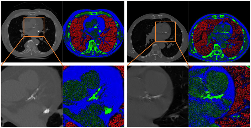
Fig. 2:
Two examples of anatomical-information based multichannel image coding. The second row is the magnification of the heart region in the first row. With the proposed coding scheme, the large intensity range of CT images can be divided into three smaller segments to highlight the important imaging features. Red areas mostly correspond to emphysema severity; blue areas represent fat attenuation concentrated regions; green areas contain mostly calcification or bone.

