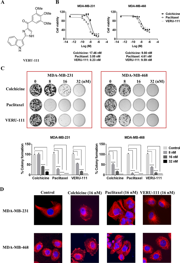
Figure 1.
Efficacy of VERU-111 versus paclitaxel in two TNBC cell lines, MDA-MB-231 and MDA-MB-468. (A) Chemical structure of VERU-111. (B) The anti-proliferative effect of VERU-111 relative to colchicine and paclitaxel controls determined by MTS assay after 72 h (response curves representative of three independent biological replicates are shown; also refer to Supplementary Table S1). (C) Representative pictures of a colony formation assay using MDA-MB-231 (left panel) and MDA-MB-468 (right panel) cells treated with colchicine, paclitaxel and VERU-111 at increasing concentrations (8, 16 or 32 nM) for 7 days and 14 days, respectively. Quantification of colony formation area is expressed as the grand mean ± SEM compared to vehicle control (set to 100%) of three biological replicate experiments. (D) Representative immunofluorescence anti-tubulin staining images of the microtubule network in MDA-MB-231 (top panel) and MDA-MB-468 (lower panel) cells treated with vehicle (Control), colchicine (16 nM), paclitaxel (16 nM) and VERU-111 (16 nM) for 16 h. Images were obtained at ×20 magnification.

