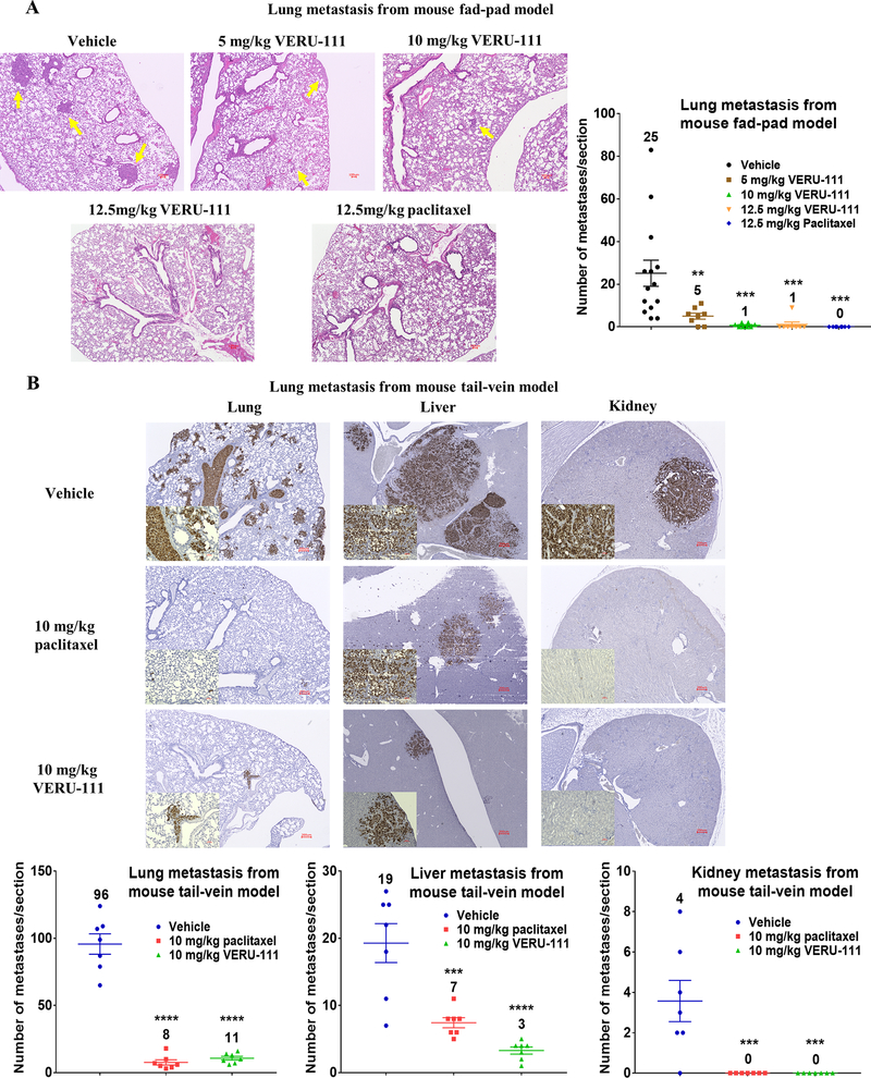
Figure 6.
VERU-111 treatment suppresses metastasis formation in an orthotopic xenograft model and in an experimental lung metastasis model using MDA-MB-231 cells. (A) Representative H&E stained sections of lung metastases derived from orthotopically implanted MDA-MB-231 cells. Lung metastases are indicated by yellow arrows in the H&E-stained slides, 4x magnification, scale bar = 100 μm. Lung metastatic burden of each section from each mouse was quantified after scanning whole slides. (B) Anti-human specific mitochondria IHC staining to detect metastases in lung, liver and kidney sections, scale bar is 200 μm for primary figures and 50 μm for inserts. Scatter plots of mean ± SEM show the quantification of metastases present in the lung, liver and kidney (A-B). The number above each treatment group correspond to the mean.

