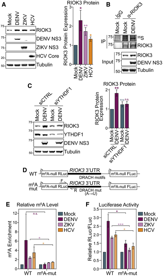
Figure 3: m6A promotes RIOK3 protein expression.
(A) (Left) Representative immunoblot of RIOK3 protein expression in mock- and virus-infected (48 hpi) Huh7 cells. (Right) Quantification of RIOK3 protein expression relative to tubulin. (B) Immunoprecipitation (IP) of RIOK3 from mock- and DENV-infected (48 hpi) Huh7 cells labeled with 35S for 3 hours. IP fractions were analyzed by autoradiography (35S) and immunoblotting. Representative of 3 biological replicates. (C) (Left) Representative immunoblot of RIOK3 protein expression in mock- and DENV-infected (48 hpi) Huh7 cells treated with non-targeting control (CTRL) or YTHDF1 siRNA. (Right) Quantification of RIOK3 protein expression relative to tubulin. (D) Schematic of WT and mutant m6A-null Renilla luciferase (RLuc) RIOK3 3’ UTR reporters that also express m6A-null Firefly luciferase (FLuc) from a separate promoter. RT-qPCR primers (F and R) are indicated with arrows. (E) MeRIP-RT-qPCR analysis of relative m6A level of stably expressed WT and m6A-mut RIOK3 3’ UTR reporter RNA in mock- and virus-infected (48 hpi) Huh7 cells. (F) Relative luciferase activity (RLuc/FLuc) in mock- and virus-infected (48 hpi) Huh7 cells stably expressing WT and m6A-mut RIOK3 3’ UTR reporters. Relative luciferase activity in uninfected cells was set as 1 for each reporter. Values are the mean ± SEM of 6 (A), 4 (C), 2 (E), or 5 (F) biological replicates. * p < 0.05, ** p < 0.01, *** p < 0.001 by unpaired Student’s t test. n.s. = not significant. See also Figure S3.

