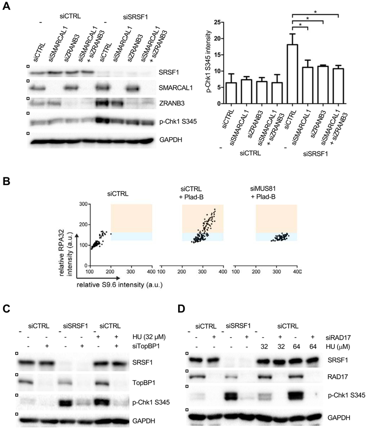
Figure 5. R loop-induced ATR activation involves replication fork reversal and MUS81-mediated ssDNA formation.
(A) HeLa cells were transfected with control, SRSF1-1, SMARCAL1, and ZRANB3 siRNA as shown. Levels of p-Chk1 and other proteins were analyzed by western blot (left panel). The levels of p-Chk1 from three independent experiments were quantified (n=3) (right panel). Error bars, SD. *p < 0.05, Unpaired Student’s t test. (B) Hela cells were transfected with control or MUS81 siRNA for 48 hr. Cells were then treated with Plad-B (1 μM) for 4 hr. Individual cells were analyzed by immunostaining using RPA32 and S9.6 antibodies. Intensities of S9.6 and RPA staining of individual cells were analyzed and plotted in 2D. (C-D) HeLa cells were transfected with control, SRSF1-1, TopBP1 (C), and RAD17 (D) siRNA as shown. Where indicated, cells were treated with 32 or 64 μM of HU for 1 hr. Levels of p-Chk1 and other proteins were analyzed by western blot.

