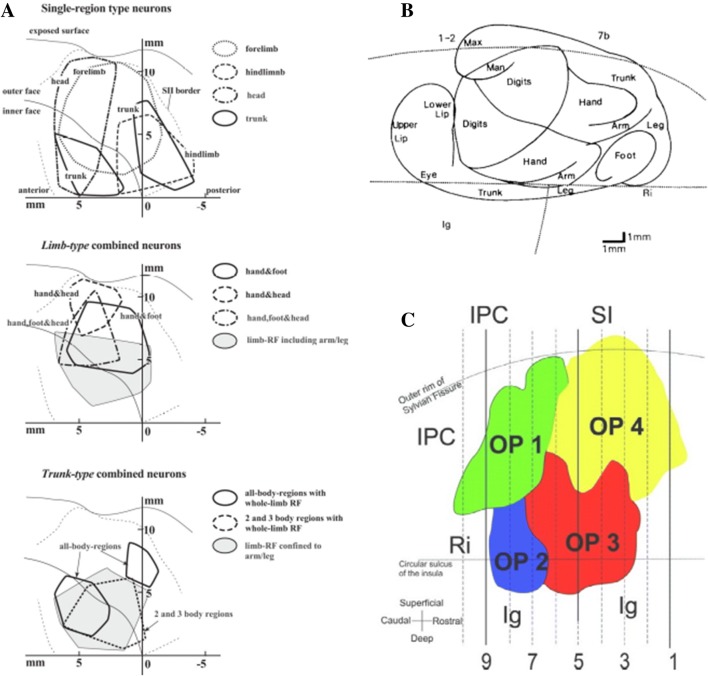
Fig. 1.
Cortical organization of the parietal operculum. a Unfolded surface of the lateral sulcus of macaque showing the mirrored distribution of somatosensory responsive neurons of different body parts (electrophysiological data). The vertical axis indicates distance from the upper fundus (Taoka et al. 2016). b Distribution in the SII of anterograde tracers injected into closely related cutaneous responsive sites in macaque (Burton et al. 1995). Same representation format as (a). c Flattened representation of the four cytoarchitectonic areas in the human parietal operculum (OP for operculum). Indicated on figure: inferior parietal cortex (IPC), retroinsula (Ri), primary somatosensory cortex (SI), and granular insular cortex (Ig). OP 4 and OP 1 are suggested to be the human analogues of the primate parietal ventral area (PV) and SII, respectively (Eickhoff et al. 2006)

