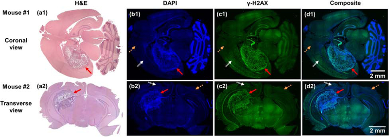
Figure 6.
(a1–c1 and a2–c2) are H&E, DAPI and γ-H2AX staining in coronal and transverse brain sections from two mice, respectively, and (d1 and d2) are the composited images of DAPI and γ-H2AX staining. Red solid, orange dash, and white double line arrows point to the GBM, normal tissue, and normal tissue irradiated area, respectively.

