
Figure 2.
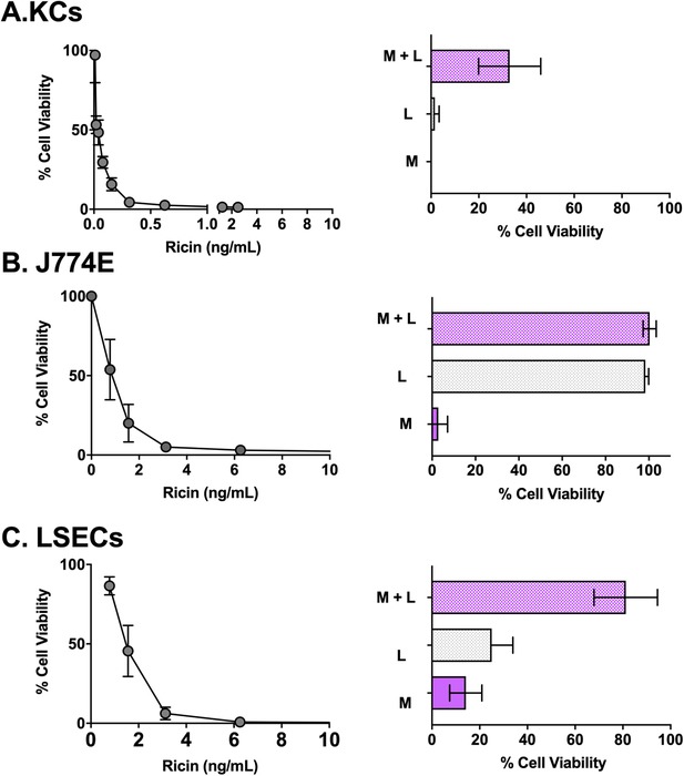
Sensitivity of KCs, J774E cells, and LSECs to ricin toxin. Left panels: Average cell viability of KCs (panel A), J774E cells (panel B), and LSECs (panel C) after an 18 h exposure to indicated concentrations of ricin toxin from 2 independent experiments. Right panels: Effects of coincubation of ricin with α‐mannan (M; 1 mg/ml), lactose (L; 0.1 M), and the combination of α‐mannan and lactose (M + L) on viability of the 3 different cell types. Each bar represents the average (with corresponding standard deviations) of 6 independent experiments