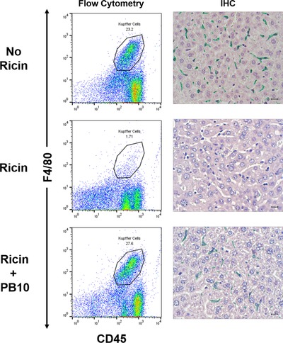
Figure 5.

PB10 protects KCs from the effects of ricin in vivo. Groups of mice were challenged by i.p. injection with saline (top row), ricin toxin (middle row), or ricin toxin plus PB10 (bottom row). Eighteen hours later, the mice were euthanized, and liver tissues collected for KC isolation (left panels) or IHC (right panel). For flow cytometric analysis, KCs were immunostained for F4/80 (y‐axis) and CD45 (x‐axis), as described in the section Materials and Methods. The CD45+ F4/80+ KC populations are encircled, and the percentage of the total cell numbers are noted. For IHC (right panels), formalin‐fixed tissue sections were stained with anti‐F4/80 followed by HRP‐based polymer and Vina Green chromogen, also as described in the section Materials and Methods. Note the complete absence of F4/80+ cells in ricin treated mice, compared to near normal numbers of F4/80+ cells in ricin plus PB10‐treated animals
