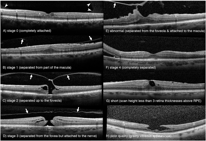
FIGURE 1.
Example 6 mm optical coherence tomography scans demonstrating posterior vitreous detachment classification. Attached vitreous includes stage 0 (A), stage 1 (B), stage 2 (C), stage 3 (D), and abnormal stage (E) and was identified by either the premacular bursa (arrowheads) or posterior vitreous cortex (arrows). Complete posterior vitreous detachment is represented by stage 4 (F), in which neither the premacular bursa nor the posterior vitreous cortex were visualized. Scans were excluded if they had insufficient scan height (G) or insufficient scan quality (H). Image brightness has been adjusted to maintain quality during printing. Staging system modified from Ma et al.12

