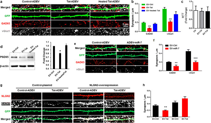
Fig. 4.
miR-7 in Tat-ADEVs causes synaptic injury. a Representative confocal images of GFP-expressing PRHNs exposed to indicated ADEVs (24 h), stained with GAD65 and vGlut1 & b quantification of excitatory & inhibitory synapses. c Cell viability was measured using MTT assay. d Representative western blot image and quantification of PSD95 in PRHNs exposed to indicated ADEVs. e Representative confocal images of GFP-expressing PRHNs exposed to ADEVs loaded with either control or miR-7 oligos (24 h) & stained with GAD65 & vGlut1 & f quantification of excitatory and inhibitory synapses. g Representative confocal images of GFP and NLGN2-expressing PRHNs exposed to the indicated ADEVs (24 h) & stained with NLGN2 & PSD95 & h quantification of puncta for each condition. One way ANOVA with post hoc test. Bars represent mean ± SD from 3 independent experiments. *p < 0.05; ** p < 0.01; *** p < 0.001vs control; # p < 0.05 vs Tat-ADEV group

