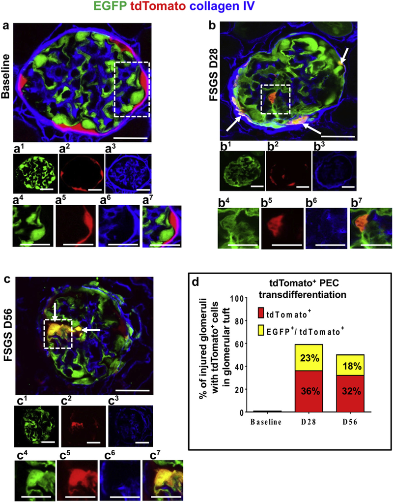
Figure 2|. Transdifferentiation of parietal epithelial cells (PECs) to a podocyte fate after podocyte depletion.

(a–c) Confocal microscopic images of enhanced green fluorescent protein (EGFP)þ podocytes (green), tdTomato+ PECs (red), podocytes derived from PEC origin (yellow), and collagen IV (blue, used to delineate kidney architecture). In panels labeled with numbers, 1 (green), 2 (red), and 3 (blue) are fluorescent channels of whole glomeruli and 4 (green), 5 (red), 6 (blue), and 7 (merged) are channels of the area outlined by the white dashed boxes. (a) At baseline, tdTomato+ PECs are limited to Bowman’s capsule and EGFP+ podocytes to the glomerular tuft, with no overlap. (b) At focal segmental glomerulosclerosis (FSGS) day 28 (D28), a subpopulation of yellow cells are detected along Bowman’s capsule (arrows) and on the glomerular tuft (within the dashed box). (c) At FSGS day 56 (D56), yellow cells (arrows) are detected on the glomerular tuft (marked with dashed box). There is an overall decrease in EGFP+ podocytes, and fewer tdTomato+PECs are detected along Bowman’s capsule. (d) Quantitative analysis of the percentage of injured glomeruli with tdTomato+ PECs in the glomerular tuft. tdTomato+ PECs (red) and PECs that transdifferentiated to a podocyte fate (yellow) were not detected in the glomerular tuft in baseline glomeruli nor in uninjured glomeruli at FSGS day 28 and day 56. Of the subset of injured glomeruli, 36% had 1 or more undifferentiated tdTomato+ PECs (red), whereas 23% of the injured glomeruli had transdifferentiated PECs (yellow) at day 28 of FSGS. At day 56, 32% of the injured glomeruli had undifferentiated tdTomato+ PECs (red) and 18% had transdifferentiated PECs (yellow). Bars = 25 μm or5 μm (insets).
