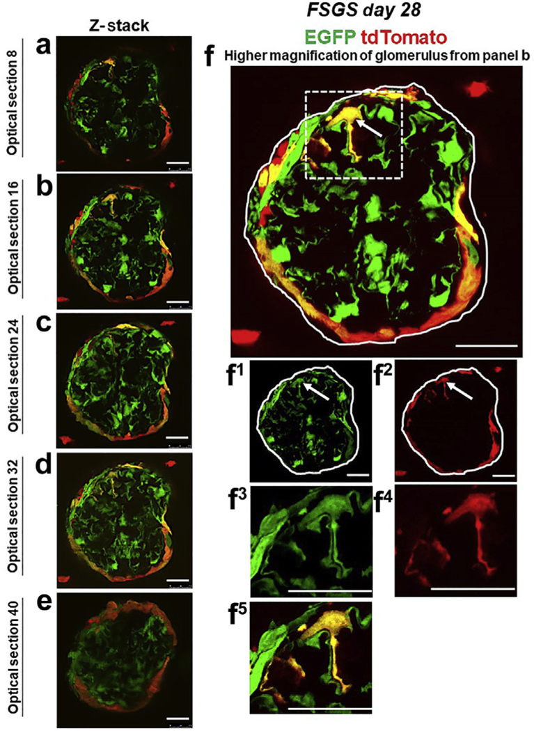
Figure 3|. tdTomato+ parietal epithelial cells (PECs) on the glomerular tuft at focal segmental glomerulosclerosis (FSGS) day 28 show genetic evidence of transdifferentiation toward a podocyte fate.

(a–f) Consecutive confocal optical images compiled as a Z-stack through a glomerulus at FSGS day 28 show enhanced green fluorescent protein (EGFP; green), tdTomato (red), and merge (yellow, solid arrows show example). Yellow cells are detected on the glomerular tuft, and the density of tdTomato+ PECs along Bowman’s capsule is substantially reduced as cells move from Bowman’s capsule to the glomerular tuft. (f) A higher magnification of optical section 16 from the Z-stack. Single fluorescent channels of the inset marked by the dashed box show EGFP (f1) and tdTomato (f2). Further magnified images of the area marked by the dashed box with a migrated tdTomato+ EGFP+ PEC are shown in following panels: (f3) EGFP, (f4) TdTomato, and (f5) merge. Antibodies were not required to detect EGFP and tdTomato reporters. These results provide genetic proof that at FSGS day 28, a subset of tdTomato+ PECs that migrated to the glomerular tuft transdifferentiate to a podocyte fate. Bars = 25 μm or 5 μm (insets).
