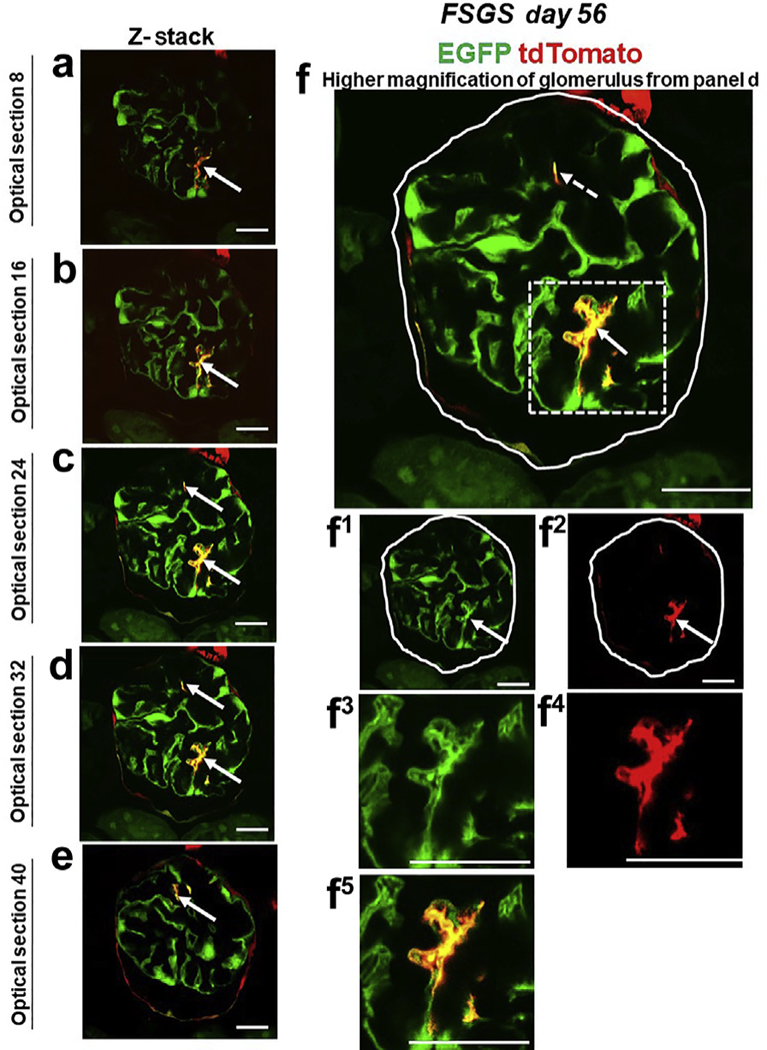
Figure 4|. tdTomato+ parietal epithelial cells (PECs) on the glomerular tuft at focal segmental glomerulosclerosis (FSGS) day 56 show genetic evidence of transdifferentiation toward a podocyte fate.

(a-e) Consecutive confocal optical images from top to bottom through a glomerulus compiled as a Z-stack of PEC-rtTA│LC1│tdTomato│Nphs1-FLPo│FRT-EGFP (PEC-PODO) mice given doxycycline at FSGS day 56 show enhanced green fluorescent protein (EGFP; green), tdTomato (red), and merge (yellow). Yellow cells are detected on the glomerular tuft and are particularly visible in panels b-d. Panels c-e show evidence of another yellow cell. (f) A higher magnification of optical section 32 from the Z-stack. Single fluorescent channels of the inset marked by the dashed box show EGFP (f1) and tdTomato (f2). Further magnified images of the area marked by the dashed box with a migrated tdTomato+ EGFP+ PEC are shown in panels (f3) EGFP, (f4) TdTomato, and (f5) merge. Antibodies were not required to detect EGFP and tdTomato reporters. These results provide genetic proof that at FSGS day 56, a subset of tdTomato+ PECs that migrated to the glomerular tuft transdifferentiate to a podocyte fate. Bars = 25 μm or 5 μm (insets).
