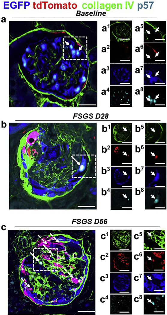
Figure 7|. A subset of tdTomato+ parietal epithelial cells (PECs) that have migrated to the tuft after podocyte loss acquire a podocyte fate and begin to express the podocyte protein p57.

Representative images of 4-color immunostaining at baseline, day 28 (D28), and day 56 (D56) of focal segmental glomerulosclerosis (FSGS). The panels in the 2 right columns show individual colors for collagen IV (green), tdTomato (red), enhanced green fluorescent protein (EGFP; blue), and p57 (cyan); the column on the far right shows higher magnification images of the area highlighted with the dashed boxes. (a) At baseline, the majority of EGFP+ cells (blue) (a3, a7) co-localized with p57 (a4, a8) and tdTomato+ PECs (red) (a2, a6) are detected along Bowman’s capsule, delineated by collagen IV (green) (a1, a5). (b) At day 28 FSGS, a tdTomato+ PEC (red) (b2, b6) has migrated to the glomerular tuft, transdifferentiated to a podocyte fate, and co-expresses EGFP (blue) (b3, b7) and p57 (cyan) (b4, b8), creating a white color (b), marked with a solid arrow. Bowman’s capsule and glomerular tuft are delineated with collagen IV (green) (b1, b5). (c) At day 56 FSGS, a number of tdTomato+ PECs (red) (c2, c6) have migrated to the glomerular tuft, transdifferentiated to a podocyte fate, and co-expressed EGFP (blue) (c3, c7; c, magenta color, dashed arrows). A subpopulation of those cells co-stained for the podocyte marker p57 (cyan) (c4, c8) (c, white color, solid arrows). Bowman’s capsule and the glomerular tuft are delineated with collagen IV (green) (c1, c5). Bars = 25 μm or 5 μm (insets).
