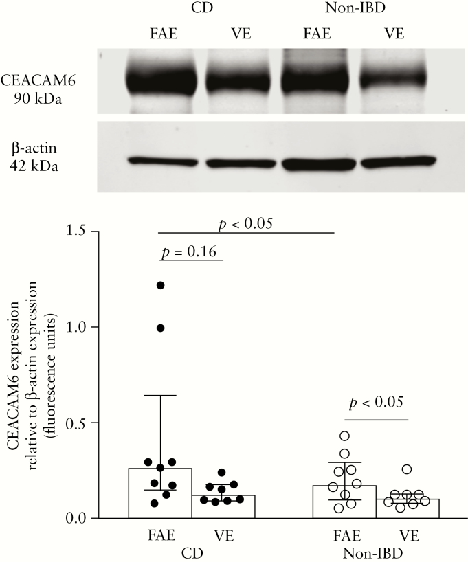
Figure 6.
Expression of CEACAM6 measured by western blotting in the follicle-associated epithelium [FAE] and villus epithelium [VE] of patients with Crohn’s disease [CD] and of non-inflammatory bowel disease [Non-IBD] controls. Segments of FAE and VE were obtained from ileum of the same individual and run in parallel. Tissue lysates from eight CD patients and eight controls were analysed in duplicate and the image shows a representative blot from one CD and one control, respectively. CEACAM6 [90 kDa] values were normalized to the loading control β-actin [mw 42 kDa]. Values represent the median [25th–75th percentiles]. Comparisons between two groups were done with the Mann–Whitney U test.

