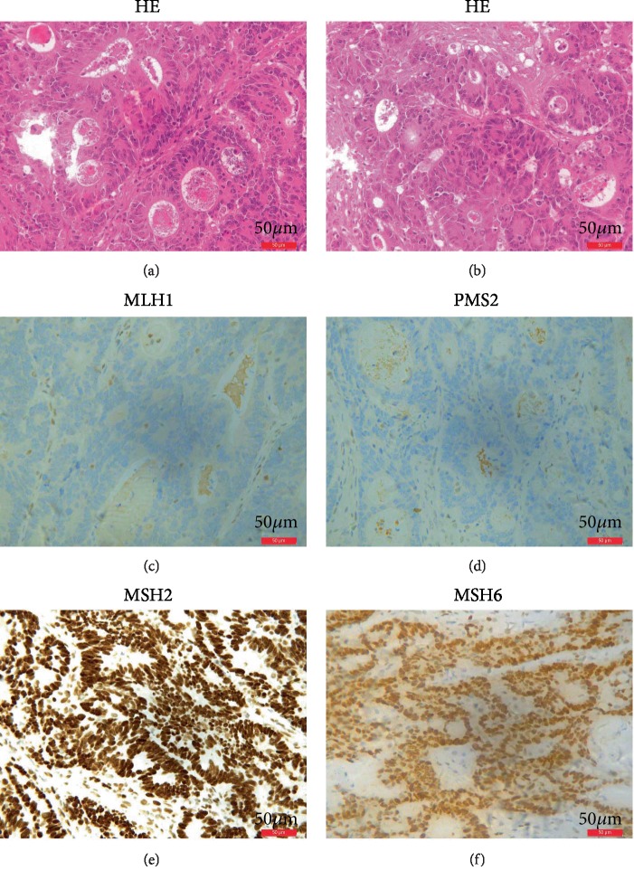
Figure 5.
HE and IHC staining results for MMR genes. (a, b) HE staining shows abnormal growth and shape of tumor cells from the proband. (c) IHC staining showed loss of expression of MLH1. (d) IHC staining showed loss of expression of PMS2. (e) IHC staining showed normal expression of MSH2. (f) IHC staining showed normal expression of MSH6.

